
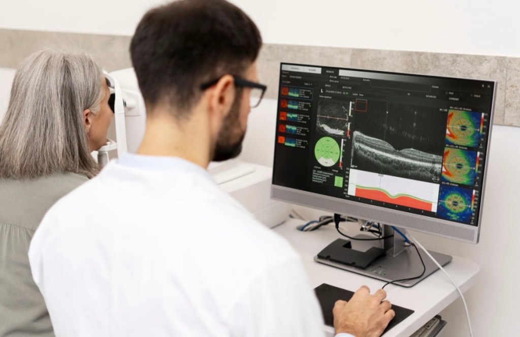
Optical Coherence Tomography (OCT) is a non-invasive imaging technique used in eye care to produce high-resolution cross-sectional images of the retina (the light-sensitive tissue at the back of the eye).
It works using light waves, similar to how ultrasound uses sound waves, to capture detailed images of the different layers of the retina. The test is quick, painless, and does not involve direct contact with the eye.
OCT allows eye specialists to see structures beneath the surface of the retina that cannot be observed during a routine eye examination.
Uses
1. Diagnosis of Retinal Diseases
OCT is widely used to detect and monitor conditions such as:
• Macular Degeneration
• Diabetic retinopathy
• Macular edema
It helps identify swelling, thinning, or damage in retinal layers.
2. Glaucoma Detection and Monitoring
OCT measures the thickness of the optic nerve fibers, which is essential in diagnosing and managing Glaucoma
3. Assessment of the Macula
It provides detailed images of the macula (central vision area), helping detect abnormalities that affect sharp vision.
4. Monitoring Treatment Progress
Doctors use OCT to track how well treatments (like injections or medications) are working over time.
5. Early Detection of Eye Problems
OCT can detect subtle changes in the retina before symptoms appear, allowing for early intervention and prevention of vision loss.
Optical Coherence Tomography is a powerful diagnostic tool that gives a detailed “inside view” of the retina. It plays a crucial role in early diagnosis, accurate monitoring, and effective treatment of many eye diseases.

Leave a comment